Gráinne's Projects

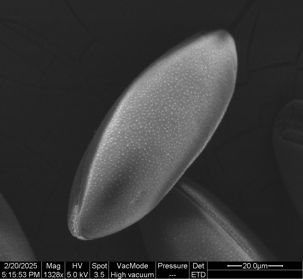
Freezing and Pollen Coat Morphology

The pollen coat has complex ornamentation that is often species-specific and may aid in identification. Freezing pollen is known to preserve its viability, but whether it preserves surface morphology had not previously been reported. In this project, I investigated whether freezing pollen affected the morphology of the pollen coat. I found that Narcissus pollen coat ornamentation remained unaffected both by freezing and by storage at room temperature for two months.

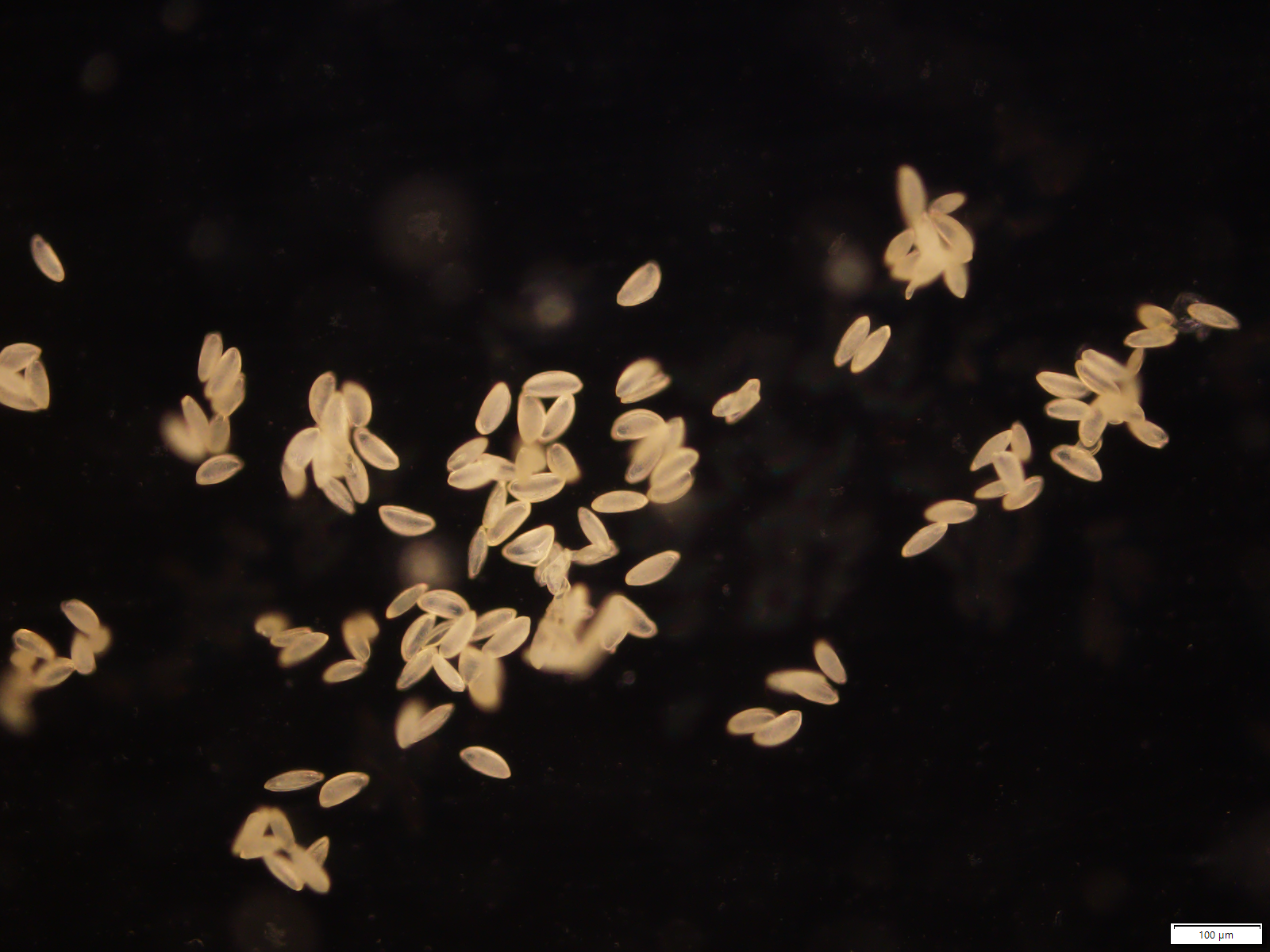
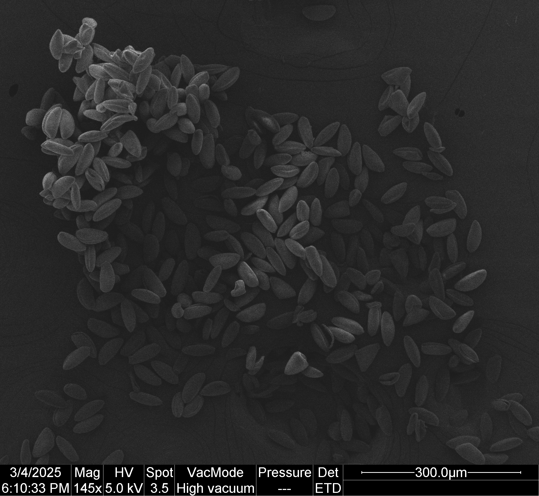
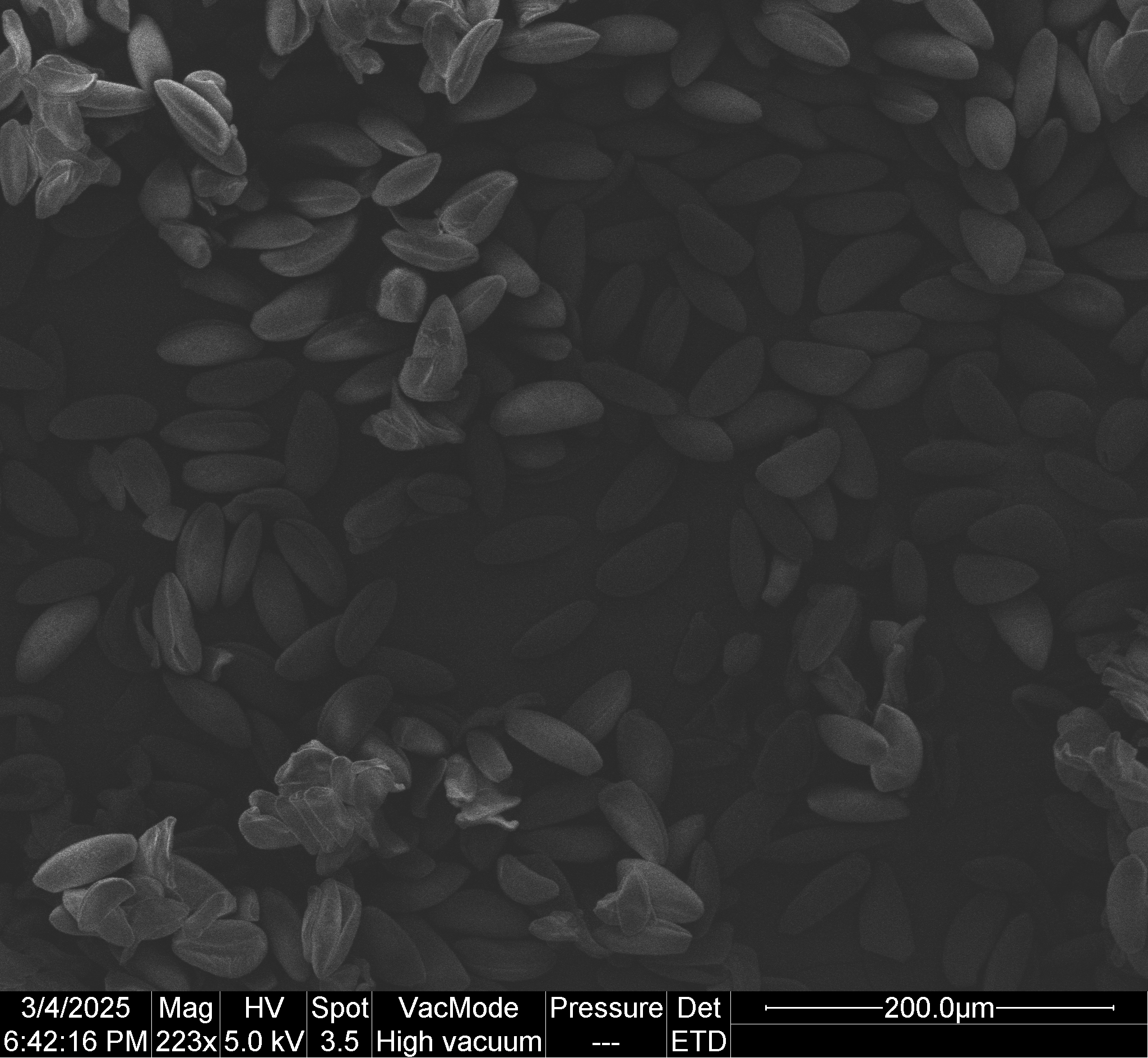
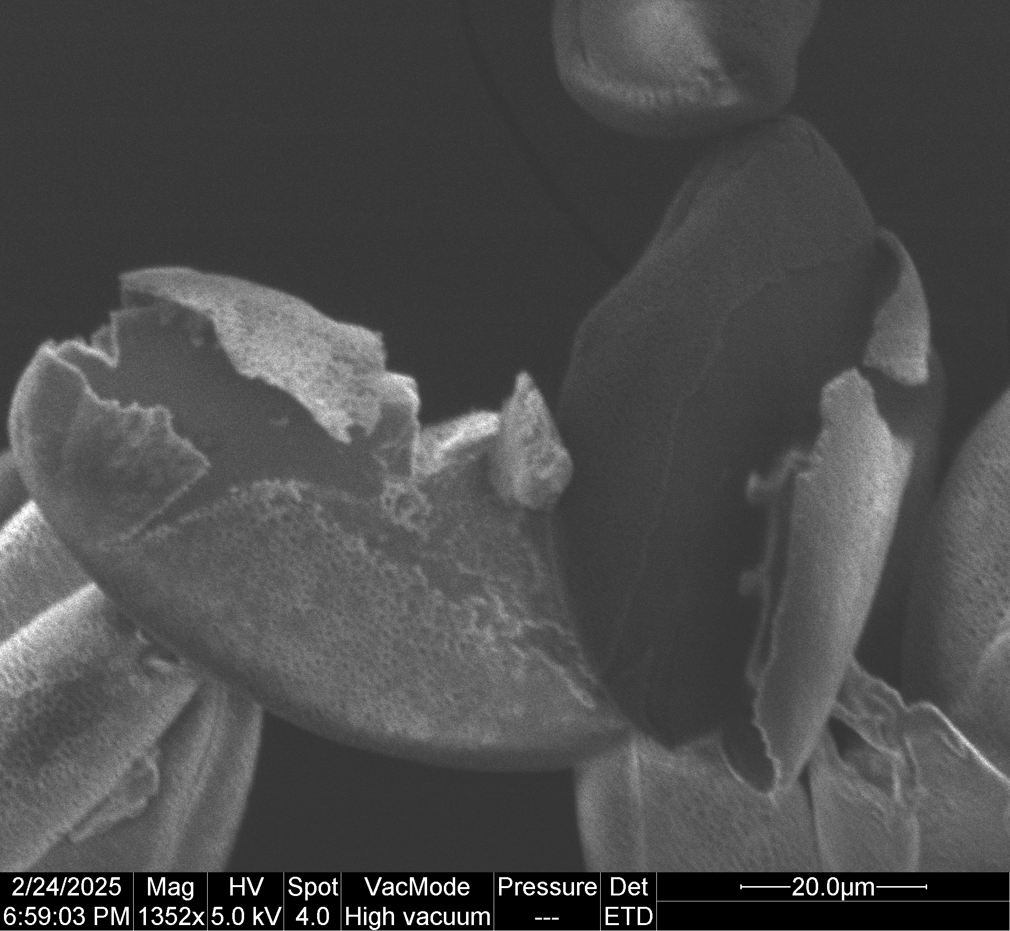
Research Micrographs


          *Narcissus* pollen grains kept at room-temperature for one week. The pollen was sputter coated in gold and imaged with a scanning electron microscope using a secondary electron detector. Scale bar indicates 300 µm.
        
          *Narcissus* pollen grains kept at -80°C for one week. The pollen was thawed, sputter coated in gold, and imaged with a scanning electron microscope using a secondary electron detector. Scale bar indicates 200 µm.
        
          *Narcissus* pollen grains kept at room-temperature for eight weeks. The pollen was rinsed with ethanol and hydrated with 50% glycerol in water before imaging. Imaging was conducted with a laser scanning confocal microscope using a laser of wavelength 405.0 nm, resulting in autofluorescence in the blue range. The micrograph shows a maximum intensity projection from images taken at 32 vertical positions. Scale bar indicates 50 µm.
        
          *Narcissus* pollen grains kept at -80°C for eight weeks. The pollen was thawed, rinsed with ethanol, and hydrated with 50% glycerol in water before imaging. Imaging was conducted with a laser scanning confocal microscope using a laser of wavelength 405.0 nm, resulting in autofluorescence in the blue range. The micrograph shows a maximum intensity projection from images taken at 32 vertical positions. Scale bar indicates 50 µm.

Other Interesting Micrographs


          A *Freesia* pollen grain, showing the surface texture of the pollen coat. The pollen was sputter coated in gold and imaged with a scanning electron microscope using a secondary electron detector. Scale bar indicates 20 µm.
        
          A fractured *Narcissus* pollen grain, with the outer layers separated from the rest of the pollen grain. The pollen was sputter coated in gold and imaged with a scanning electron microscope using a secondary electron detector. Scale bar indicates 20 µm.

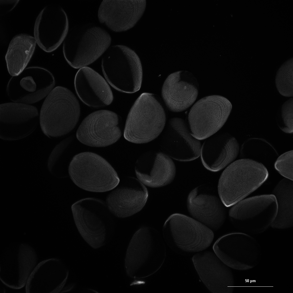
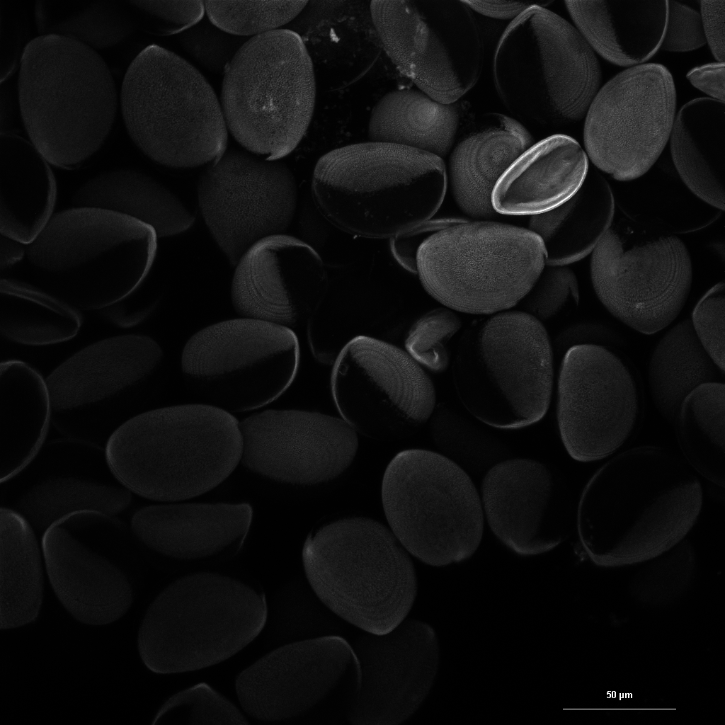
*Narcissus* pollen grains in whole mount. The pollen was rinsed with ethanol and hydrated with 50% glycerol in water before imaging. Imaging was conducted with a laser scanning confocal microscope using a laser of wavelength 405.0 nm, resulting in autofluorescence in the blue range, and a laser of wavelength 490.1 nm, resulting in autofluorescence in the green range. The micrograph shows a 3D volume rendering from images taken at 32 vertical positions, false colored to indicate emission wavelength. The imaged area is approximately 212×212×33 µm in size.


          *Narcissus* pollen grains in whole mount. The pollen was observed dry using a compound microscope with reflected light darkfield illumination. Scale bar indicates 100 µm.
        
          Fractured *Narcissus* pollen grains in whole mount, showing the separation of different layers of the pollen grains. The pollen was observed dry using a compound microscope with reflected light darkfield illumination. Scale bar indicates 20 µm.